Laboratory of Molecular Biology, Biochemistry and Biophysics (LMBBB) is dedicated to research and teaching activities.
The main research facilities include equipment designed for basic and advanced biochemical and biophysical analyses of microbial, plant and animal cell compounds. Separation, quantitative and qualitative measurements of proteins, carbohydrates, lipids and nucleic acids can be performed at our premises employing spectrophotometers, spectrofluorimeter, chromatographic and gel-electrophoresis units. Molecular investigations of genotypic and phenotypic features of living cells can be achieved by PCR, IEF, Western blotting and protein array techniques. Additionally, animal and cell cultures facilities and expertise are available at the premises of our research laboratory.
Research interests/fields:
- biomedical biochemistry;
- bionanomedicine;
- environmental molecular biology;
- molecular diversity and taxonomy;
- cell physiology.
Domains of activity
- Animal cell culture:
- Genomics, proteomics and NGS data analysis;
- Microbial cell culture;
- Biochemical analyses.
Staff
General biochemistry, immunobiology and bionanotechnology
- Assoc. Prof. Manuela BANCIU, PhD
- Lecturer Emilia LICĂRETE, PhD
- Lecturer Alina SESĂRMAN, PhD
- Teaching assistant Laura PĂTRAȘ, PhD student
Biophysics, bioinformatics, molecular enviornmental microbiology
- Prof. Horia BANCIU
- Teaching assistant Paul-Adrian BULZU, PhD
Major infrastructure
Guava® Muse® flow microcytometry system for cell culture characterization (Cytek Bioscience)
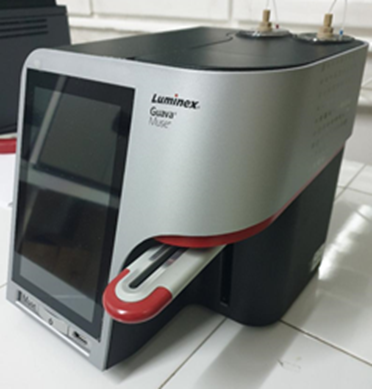
Specifications: The Guava® Muse® Cell Analyzer is a compact and portable instrument for fluorescence-based analysis, consisting of cell analysis software and reagents optimized for performing a wide range of cell assays with up to two fluorescent parameters in addition to direct scatter determination. The device is designed for easy and intuitive use and minimal maintenance. The integrated touch screen provides an easy-to-use software interface for operating the system. The Guava® Muse® Cell Analyzer uses miniaturized fluorescent detection and microcapillary technology to provide quantitative cell analysis of both suspended and adherent cells from 2 to 60 μm in diameter. The software includes a dedicated module for each test, as well as a tool for checking performance and cleaning the instrument’s fluid system. The modules allow researchers to stain samples with their own antibodies conjugated to fluorochromes, dyes, or other reagents that are excited by a 532 nm laser. The yellow parameter uses a 576/28 emission detection channel and can be used to detect fluorochromes such as Phycoerythrin (PE), Cy3, Alexa Fluor® 555 and Dylight® 550. The red parameter uses a 680 emission detection channel /30 and can be used for the detection of fluorochromes such as PE-Cy5, 7-AAD and propidium iodide (PI).
Applicability: cell counting, viability assays, proliferation, apoptosis and oxidative stress, determination of cell cycle phases, study of extracellular and intracellular expression of fluorescently labeled receptors
Fields: molecular biology, molecular diagnosis, immunology, microbiology, biochemistry, medical biology
Teaching provided: Metabolic biochemistry, Bionanotechnologies; Biochemistry of proteins and introduction to proteomics, Biochemistry of nucleic acids and introduction to genomics; Immunobiology, Medical Biochemistry, Modern Biochemical and Biophysical Methods, (3) Immunobiology; Environmental molecular biology, at the Bachelor’s degree specializations (Biochemistry, Biology and Industrial Biotechnologies) and Master (Molecular Biotechnology and Medical Biology (LR and LM); PhD – Doctoral School of Integrative Biology) specializations. Person in-charge: Lecturer dr. Rauca Valentin-Florian – valentin.rauca@ubbcluj.ro; Lecturer dr. Pătraș Laura-Ioana laura.patras@ubbcluj.ro.
Access: Mo to Fri, 8-18; with e-mail programming.
Axio Vert.A1 FL inverted microscope with LED fluorescence source and AxioCam 208c color digital camera (Carl Zeiss, Germany)
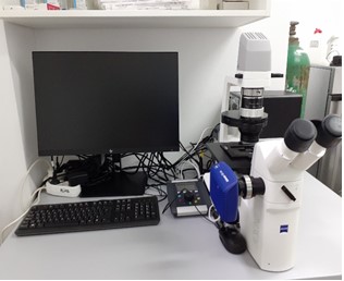
Specifications: Axio Vert.A1 FL inverted microscope (Carl Zeiss Instruments, Germany) for examination in transmitted light, light field and phase contrast. State-of-the-art LED fluorescence source. AxioCam 208c color digital camera. Image acquisition and analysis software.
Uniqueness: The inverted microscope with state-of-the-art fluorescence source – Colibri 5, has 4-channel LED for viewing the following fluorochromes: Red: at 630nm, for Cy5 excitations, Alexa 631 nm, Toto-3 and similar colors; Green: at 475 nm, for Cy3, TRITC, DsRED excitations and similar colors; Blue: at 475nm for eGFP, Fluo4, FITC excitations and similar colors; Ultraviolet: at 385 nm for DAPI, Alexa 405, Hoechst 33258 and similar colors. It has the option of generating high resolution images with overlapping colors. The microscope has ECO Power function, a 8.3 Megapixel AxioCam 208c digital camera and image acquisition and analysis software (ZEN 3.0 lite + ZEN lite / starter Module Measurement).
Applicability: cell counting, observation of mammalian cell morphology in 2D and 3D culture, intracellular uptake studies for different fluorescent compounds, cell apoptosis studies, medical diagnostic methods (immunofluorescence), examination of biological fluids or other tissues (from plants, animals, human).Fields: Cell Biology, Immunology, Oncobiology, Hematology, Parasitology, Histopathology, Embryology, Plant/Animal/ Human Histology.
Teaching provided: Immunobiology, Modern biochemical and biophysical methods, Bionanotechnologies (Master, Molecular biotechnologies); Environmental molecular biology (Doctoral School of Integrative Biology).
Persons in-charge:
Associate Prof.Dr Habil.. Manuela Banciu – manuela.banciu@ubbcluj.ro
Lecturer Dr. Alina Sesărman – alina.sesarman@ubbcluj.ro
Lecturer Dr. Emilia Licărete – emilia_licarete@yahoo.com
Access: Mo to Fri, 8-17; with e-mail programming addressed to one of the specialists listed above.
FLUOstar Omega Filter-based multi-mode microplate ELISA reader (BMG Labtech, Germany)
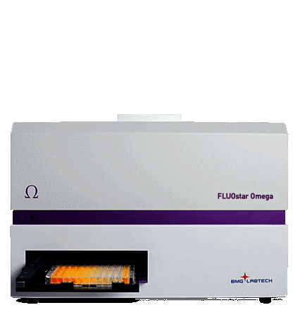
Specifications: The FLUOstar Omega multi-detection microplate reader provides the following detection modes: Ultra-fast UV/vis absorbance spectra or filter-based absorbance; fluorescence intensity (allows the use of multi-excitation and multi-emission applications, such as FRET, BRET, FURA-2 and other ratiometric methods); luminescence (flash & glow).
Uniqueness: The FLUOstar® Omega is is built upon BMG LABTECH’s unique Tandem Technology. This is a combination of two technological concepts – an ultra-fast, full spectrum absorbance spectrometer, and extremely sensitive filter-based detection, with advanced optics and a photomultiplier tube to provide superior sensitivity for all detection modes.
Applicability: detection and quantitation of antibodies against clinically relevant molecular markers, quatitation of endotoxins, nucleic acids, proteins, measurement of enzyme activity, ATP quantitation, microbial and animal cell growth monitoring by optical density.
Fields: molecular biology, biochemistry, molecular diagnostics, immunology, microbiology.
Teaching provided: Metabolic Biochemistry, Immunobiology, Biochemistry of Proteins and Introduction to Proteomics, Biochemistry of Lipids and Carbohydrates (undergraduate – Biology, Biochemistry); Modern Biochemical and Biophysical Methods, Bionanotechnology (Master – Molecular Biotechnology), Medical Biochemistry, Molecular Immunology (Master – Medical Biology)
Persons in-charge:
- Assoc Prof. Manuela Banciu – manuela.banciu[a]ubbcluj.ro
- Lecturer Dr. Alina Sesărman – alina.sesarman[a]ubbcluj.ro
- Lecturer Dr. Emilia Licărete – emilia.licarete[a]yahoo.com
Access: Mo to Fri, 8-17; with e-mail programming addressed to one of the specialists listed above.
ChemiDocTM Imaging System (Bio-Rad, USA)
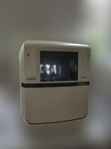
Specifications: The ChemiDoc Imaging System provides fast, reliable, and sensitive imaging and documentation of gels and chemiluminescence Western blots. This system is compatible with stain-free technology, chemiluminescence detection, and a wide range of gel stains such as ethidium bromide, SYPRO Ruby, Coomassie, and silver stains. The ChemiDoc Imaging System can be upgraded to gain the full multiplex fluorescent Western blotting capabilities
Applicability: nucleic acid and protein research.
Fields: molecular biology, proteomics, immunology, genetics, molecular diagnosis.
Teaching provided: Metabolic biochemistry; Biochemistry of proteins; Biochemistry of nucleic acids, Immunobiology (undergraduate – Biochemistry); Recombinant DNA Technologies, Medical Biochemistry (Master – Molecular Biotechnology and Medical Biology); Environmental Molecular Biology (PhD – Doctoral School of Integrative Biology).
Person in-charge: Lecturer Dr. Alina Sesărman – alina.sesarman[a]ubbcluj.ro
Access: Mo to Fri, 8-17; with e-mail programming.
Optical microscope model Axio Scope A1 with fluorescence module and color Axio Cam 105 camera (Carl Zeiss, Germany)

Specifications: Axio Scope A1 Microscope HAL 50, FL-LED, 3x H Microscope, 3x DIC with Fluorescence Module (365 nm and 540 nm fluorescence), Axio Cam 105 color camera with CMOS sensor, 5 Mpixels resolution
Uniqueness: Optical microscope with multiple functions (direct light, phase contrast, dark field, fluorescence)
Applicability: cell count, cell morphology, cellular structures (fungal, animal and plant cells), cell viability assessment.
Fields: environmental and medical microbiology, cell biology, animal cell cultures, hematology, animal / plant histology, histopathology.
Teaching provided: Biophysics (undergraduate – Biology, Biochemistry); Modern Biochemical and Biophysical Methods (Master – Molecular Biotechnology)
Persons in-charge:
- Prof. Horia Banciu – horia.banciu[a]ubbcluj.ro
- Lecturer Dr. Emilia Licărete – emilia_licarete[a]yahoo.com
- Teaching Assist. Laura Pătraș – patras.laura88[a]yahoo.com
Access: Mo to Fri, 8-17; with e-mail programming addressed to one of the specialists listed above.
CFX96 Touch™ Real-Time PCR Detection System (Bio-Rad, USA)
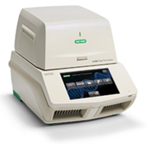
Specifications: C1000 Touch ™ thermocycler model with CFX96 ™ optical module (Bio-Rad, Hercules, CA, USA). Optical detection: Excitation 6 filtered LEDs; Detection of 6 filtered photodiodes; Range of excitation / emission wavelengths 450-730 nm; Detection sensitivity 1 copy of the target genomic DNA sequence; dedicated software for data analysis.
Applicability: amplification and quantification of gene sequences, melting curve analysis, gene expression analysis, allelic discrimination.
Fields: molecular biology, genetics, molecular diagnosis, genomics, transcriptomics.
Teaching provided: Biochemistry of nucleic acids with genomic elements (undergraduate – Biochemistry); Structure and evolution of the genome (Master – Molecular Biotechnology); Environmental Molecular Biology (PhD – Doctoral School of Integrative Biology).
Persons in-charge:
- Prof. Horia Banciu – horia.banciu[a]ubbcluj.ro
- Lecturer Dr. Alina Sesărman – alina.sesarman[a]ubbcluj.ro
Access: Mo to Fri, 8-17; with e-mail programming addressed to one of the specialists listed above.
Server System Intel R1304WFTYS WOLF PASS (Intel, USA)
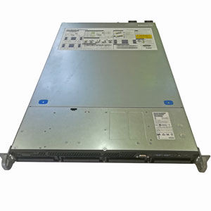
Specifications: Processor Xeon® Gold 6252 24C/48T 150W 2.10G 35.75M 10.40GT/sec ITT, Memory 12 X 64GB_LRDIMM_2666
Applicability: NGS data analysis, genome assembly, phylogenomics, comparative genomics, metabolic reconstruction
Fields: advanced bioinformatics, molecular biology, molecular ecology, biodiversity, medical genomics and proteomics, interactomics.
Teaching provided: Biochemistry of nucleic acids with genomic elements & Introduction to Bioinformatics (undergraduate – Biochemistry); Structure and evolution of the genome & Bioinformatics (Master – Molecular Biotechnology); Environmental Molecular Biology (PhD – Doctoral School of Integrative Biology).
Persons in-charge:
- Prof. Horia Banciu – horia.banciu[a]ubbcluj.ro
- Teaching Assist. Dr. Paul-Adrian Bulzu – bulzupaul[a]gmail.com
Access: Mo to Fri, 8-17; with e-mail programming addressed to one of the specialists listed above. Remote access of the server is possible upon providing the appropriate username and password.